Static visual field testing is an advanced eye examination used to measure how well you can see across your entire field of vision, including your central and peripheral sight.
In Mississauga, many eye care professionals use this test to detect early signs of eye conditions such as glaucoma, optic nerve disorders, or retinal diseases.
Because many of these conditions develop gradually and without pain, visual field testing plays an essential role in preserving long-term vision health.
During the procedure, patients are asked to focus on a central target while small lights of varying brightness appear in different areas of their visual field. Each time a light is seen, the patient presses a button, allowing the instrument to map areas of normal and reduced vision.
The test is quick, comfortable, and non-invasive, usually taking just a few minutes per eye.
The results provide valuable information about how the eyes and brain are processing visual information. If areas of decreased sensitivity are found, they may indicate the presence or progression of an eye disease. Detecting these changes early allows for timely treatment, which can help slow or prevent further vision loss.
In Mississauga, static visual field testing is commonly offered as part of a comprehensive eye exam. Regular testing helps ensure that subtle vision changes are detected as early as possible.
Static Visual Field Testing in Mississauga | Vision Clarity
Protect your eyesight with Vision Clarity’s static visual field testing, a quick, comfortable, and highly accurate way to detect early signs of glaucoma, retinal disease, or optic nerve damage.
Our advanced diagnostic equipment ensures precise results and personalized care for every patient.
Early detection means better protection and long-term eye health.
✅ Comprehensive eye exams
✅ Advanced visual field testing technology
✅ Fast, painless, and reliable results
📞 Call Vision Clarity today: +12898050019
Principles and Purpose of Static Visual Field Testing
Static visual field testing is an essential part of a comprehensive eye exam that helps measure how well you can see across your entire field of vision, both central and peripheral. This test allows eye doctors, optometrists, and ophthalmologists to detect areas of vision loss that may not be noticeable to the patient, especially in the early stages of glaucoma and other eye diseases.
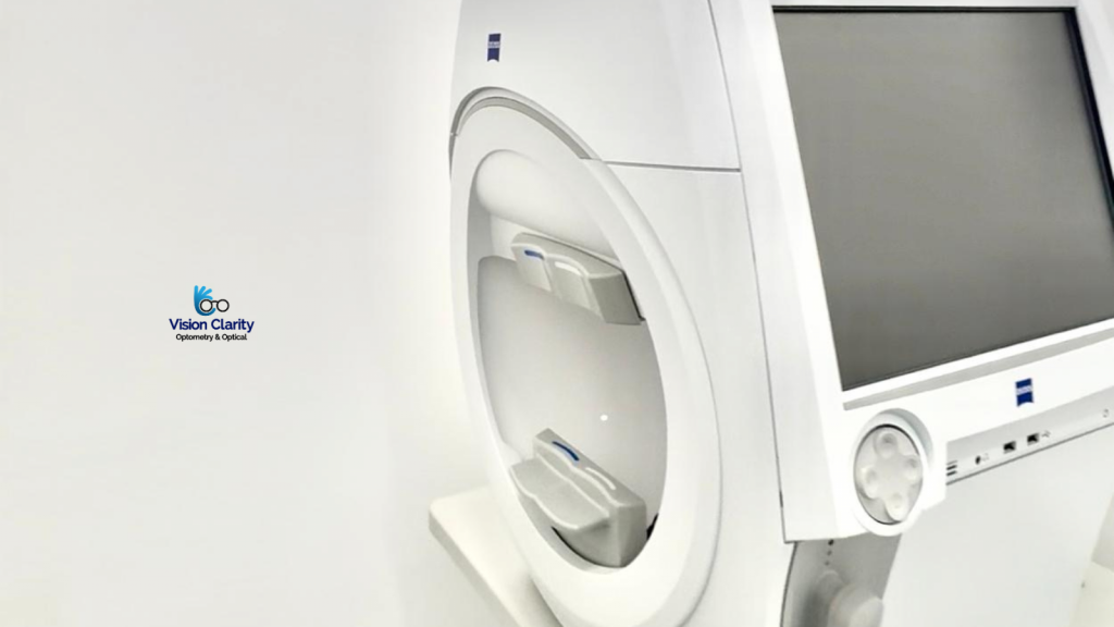
The principle behind static visual field testing is straightforward. While you focus on a central target, a series of small lights appear in different parts of your visual field. Each light varies in brightness. You simply press a button whenever you see one. The testing device, often a Humphrey Field Analyzer or similar visual field machine, records your responses to create a detailed “map” showing the sensitivity of your vision at different points.
This visual field analyzer measures the faintest light you can detect, helping your eye care professional determine the threshold sensitivity of various parts of your vision. Areas where you do not see the light may indicate potential optic nerve damage, retinal disease, or neurological conditions affecting the brain’s visual pathways.
The main purpose of static visual field testing is to detect, diagnose, and monitor eye conditions such as glaucoma, optic neuropathy, retinal detachment, and even brain tumors or strokes that may affect vision. Early detection through this test allows for timely treatment and helps prevent irreversible vision loss.
Static visual field testing is quick, painless, and non-invasive, making it a reliable way to assess eye health over time. Regular glaucoma testing and visual field exams are recommended, especially for patients over 40 or those with a family history of eye disease. By incorporating visual field testing into routine eye care, you and your eye doctor can track changes in vision and protect your sight for the future.
Equipment and Techniques Used in Static Perimetry
Static perimetry, also known as static visual field testing, is a precise and widely used method to evaluate a person’s visual field, the entire area visible while the eye is focused on a central point. This test helps eye doctors, optometrists, and ophthalmologists detect early signs of glaucoma, optic nerve damage, and other visual pathway disorders. Understanding the equipment and techniques used in static perimetry helps patients appreciate how this advanced testing supports accurate eye care.
The main equipment used for static perimetry is the automated perimeter, a computerized device designed to present stationary light stimuli at different locations within the visual field. The most common models include the Humphrey Field Analyzer (HFA), Octopus perimeter, and Medmont perimeter. These instruments feature a concave testing bowl, where the patient places their head and focuses on a central fixation point while tiny lights of varying brightness appear across the screen. The patient presses a response button whenever a light is seen.
The technique involves measuring the smallest or dimmest light stimulus that can be detected at each point, known as the threshold value. The perimeter automatically adjusts the brightness of the lights to determine this threshold, producing a detailed visual field map that shows normal and reduced sensitivity areas. Various testing strategies are available, such as Full Threshold, SITA Standard, and SITA Fast, which balance test speed and accuracy depending on the patient’s needs.
Static perimetry focuses primarily on the central 24 to 30 degrees of vision, where early signs of glaucoma often appear. Advanced versions can also test wider or specific visual zones. The data are compared against age-matched norms, allowing precise detection of subtle changes over time.
Because it is non-invasive, accurate, and repeatable, static perimetry is an essential part of modern glaucoma testing and comprehensive eye exams. Regular testing ensures early detection of vision changes, guiding treatment and helping to preserve long-term eye health.
Interpretation of Static Visual Field Test Results
Static visual field testing is a key tool for assessing the sensitivity of different areas of a person’s vision. Once the test is completed, the next crucial step is the interpretation of the results, which helps eye doctors identify early signs of eye diseases such as glaucoma, optic neuropathy, or retinal disorders. Understanding what these results mean can give patients valuable insight into their eye health and visual function.
The results of a static perimetry test are displayed as a visual field map, showing how well you can detect light stimuli at different points in your field of vision. Each point represents a specific area of the retina and is assigned a sensitivity value based on how dim a light you could see. Areas with reduced sensitivity appear as darker spots, known as visual field defects. These can indicate potential damage to the optic nerve or other parts of the visual pathway.
Eye care professionals interpret the results by analyzing several key components, including the threshold values, deviation plots, and reliability indices. Deviation plots compare your results to those of a normal, age-matched population, highlighting regions where vision may be weaker. Reliability indices help determine if the test results are accurate, for example, by tracking fixation losses or false responses during testing.
The pattern and location of any defects provide important diagnostic clues. For instance, loss of peripheral vision may suggest glaucoma, while central field defects can indicate macular or neurological conditions. Regular visual field testing allows your eye doctor to track changes over time and assess whether treatments are effectively preserving your sight.
In summary, interpreting static visual field test results requires both advanced technology and clinical expertise. By analyzing these results carefully, optometrists and ophthalmologists can detect early vision changes, guide treatment decisions, and help maintain long-term eye health and quality of vision.
Clinical Applications and Limitations of Static Visual Field Testing
Static visual field testing is a valuable diagnostic tool used by optometrists and ophthalmologists to assess the sensitivity and function of a patient’s visual field. By mapping how well a person can detect stationary light stimuli at various points, this test helps identify abnormalities in the optic nerve, retina, and visual pathways. While it plays a critical role in clinical eye care, it also has certain limitations that should be considered during interpretation.
The clinical applications of static visual field testing are extensive. One of its most common uses is in the early detection and monitoring of glaucoma, a progressive eye disease that affects peripheral vision. The test can reveal subtle areas of vision loss before noticeable symptoms appear, allowing for timely treatment and prevention of further damage. It is also widely used to evaluate optic neuropathies, retinal conditions, and neurological disorders such as stroke, pituitary tumors, or brain injuries that can affect vision.
In addition, static perimetry is useful in assessing the functional impact of eye diseases and monitoring the effectiveness of treatments over time. By comparing visual field test results from multiple visits, eye doctors can determine whether a condition is stable or progressing. This makes static perimetry an essential part of ongoing glaucoma management and comprehensive eye exams.
However, static visual field testing does have its limitations. The accuracy of results depends on patient cooperation and attention, as the test requires consistent fixation and prompt responses to visual stimuli. Fatigue, misunderstanding of instructions, or poor test reliability can affect the outcome. Furthermore, the test may not detect very small or atypical visual field defects, and it provides limited structural information compared to imaging technologies like OCT (Optical Coherence Tomography).
Despite these limitations, static visual field testing remains one of the most reliable, non-invasive, and widely accepted methods for assessing functional vision loss. When combined with other diagnostic tools, it provides a comprehensive understanding of a patient’s eye health and visual performance.